Figure 3.
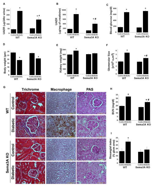
Sema3A mutant (KO) mice are resistant to diabetes induced albuminuria. Sema3A mutant mice are in C3H background. A and B: Albumin excretion rate expressed as μg/24hr urine and μg/mg of creatinine, respectively. Diabetes induced a large increase in albuminuria in WT mice which was reduced significantly in sema3A mutant mice. *, p<0.001 vs. control. #, p<0.001 vs. WT diabetic mice. C. Blood glucose in WT and sema3A mutant control and diabetic mice. *, p<0.0001 vs. control. D. Diabetes significantly reduced body weight in both WT and sema3A mutant mice. *, p<0.05 vs. control. n=8–10. E. Diabetes induced kidney hypertrophy is significantly reduced in sema3A mutant mice. *, p<0.001 vs. control. #, p<0.001 vs. WT diabetic mice. n=8–10. F. Diabetes induced a significant increase in glomerular area in WT mice which was reduced in sema3A mutant mice with diabetes. *, p<0.05 vs. control. #, p<0.05 vs. WT diabetic mice. G. Diabetes induced glomerularsclerosis and macrophage infiltration in WT and sema3A knockout mice. Trichrome staining shows fibrosis of glomeruli and the interstitium of WT diabetic mice kidney which was drastically reduced in sema3A knockout diabetic mice kidney. Similarly, extensive macrophage infiltration was seen in WT diabetic kidney as compared to control kidney. Macrophage infiltration is largely absent in sema3A knockout diabetic kidney. PAS stained section showing expansion of glomerular area, increase cellularity and deposition of proteinaceous materials in WT diabetic glomeruli which are reduced in sema3A mutant kidney section. Scale bar: 100 μM. H. Diabetes induced a significant increase in BUN in WT mice as compared to control, which was significantly reduced in sema3A KO diabetic mice. *, p<0.01 vs. control. #, p<0.05 vs. WT diabetic mice. n=6–8. I. Diabetes induced a significant increase in mesangial matrix expansion in WT mice as compared sema3A mutant mice. *, p<0.05 vs. other groups. n=8.

