
Figure 2.
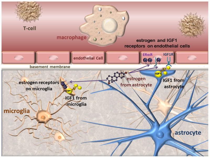
Schematic representation of the blood brain barrier: The blood brain barrier consists of closely arranged endothelial cells surrounded by basement membrane.Circulating immune cells are shown in the lumen of microvessels, while microglia are located in the brain parenchyma. Astrocytic endfeet contact the endothelial layer and provide physical and growth factor-mediated support to the barrier. Estrogen receptors and the IGF-1 receptor are located on endothelial cells, astrocytes and microglia. Astrocytes also synthesize IGF-1 and neurosteroids including estrogen. Activated microglia can also synthesize IGF-1. In cerebral ischemia, this morphological arrangement ensures steroid and growth factor support for endothelial cells.