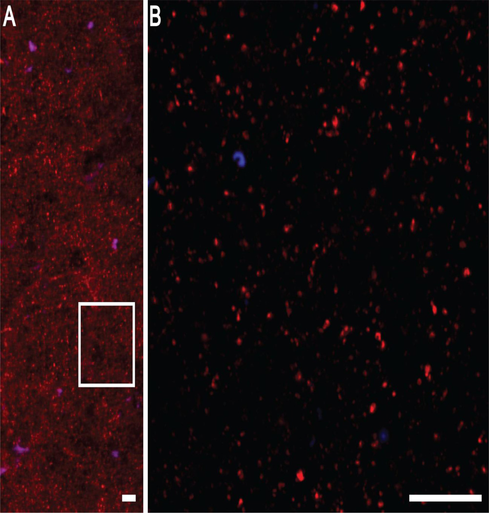
Figure 3.
Representative images of the GAD65 immunolabeling (red) used to quantify relative protein levels. A) An image collected at 10× magnification showing the predominantly punctate pattern of GAD65 labeling. B) A 60× magnification image of the boxed region in A. GAD65 labeling is confined to small, discrete punctate structures, presumed axonal boutons, in agreement with previous reports. Lipofuscin autofluorescence (blue) was predominately identified in larger structures, presumed cell bodies. Scale bars equal 10 µm.

