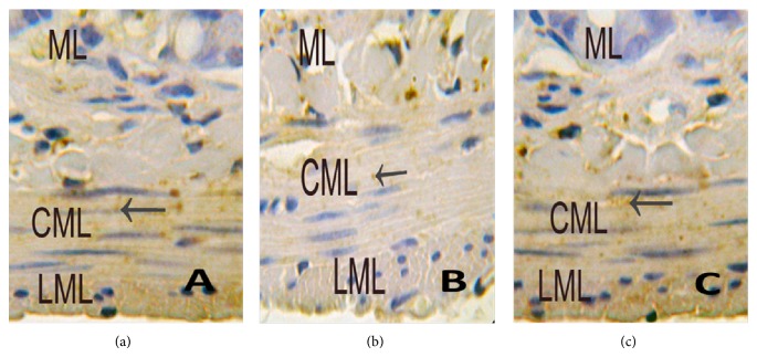
Figure 10.
Immunohistochemistry of Cx43 using light microscopy. (a) Control group shows immunoreactive products of Cx43 (←) were tan and mainly located evenly in circular muscularis, little in longitudinal muscularis, and none in mucous layer (at magnification of 400 times); (b) MODS group shows immunoreactive products of Cx43 (←) were tan and mainly located in circular muscularis, less than control group, none in longitudinal muscularis and mucous layer (at magnification of 400 times); (c) DCQD group shows immunoreactive products of Cx43 (←) were tan and mainly located evenly in circular muscularis, and there was no significant difference, compared with control group (at magnification of 400 times).

