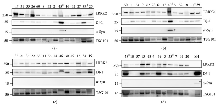
Figure 2.
Western blot results of all exosome samples. Exosome samples isolated from PD and non-PD urine samples were loaded as age- and gender-matched PD and non-PD sample pairs side by side on SDS-protein gels and analyzed by Western blotting with LRRK2, DJ-1, α-synuclein (α-syn), and TSG101 antibodies under the same conditions at the same time. The membrane was cut to the proper size and each membrane was blotted with the indicated antibody. Molecular weight markers are indicated on the left side of the blots. SH and SH-SY5Y cell lysates were used as a positive control; ‡ samples excluded from analysis because the urinalysis showed presence of protein; * monomeric form of α-synuclein. Numbers ≤ 31 indicate PD and numbers ≥ 32 indicate non-PD control samples. The male samples are shown in gels (a) and (b), and the female samples are shown in gels (c) and (d).

