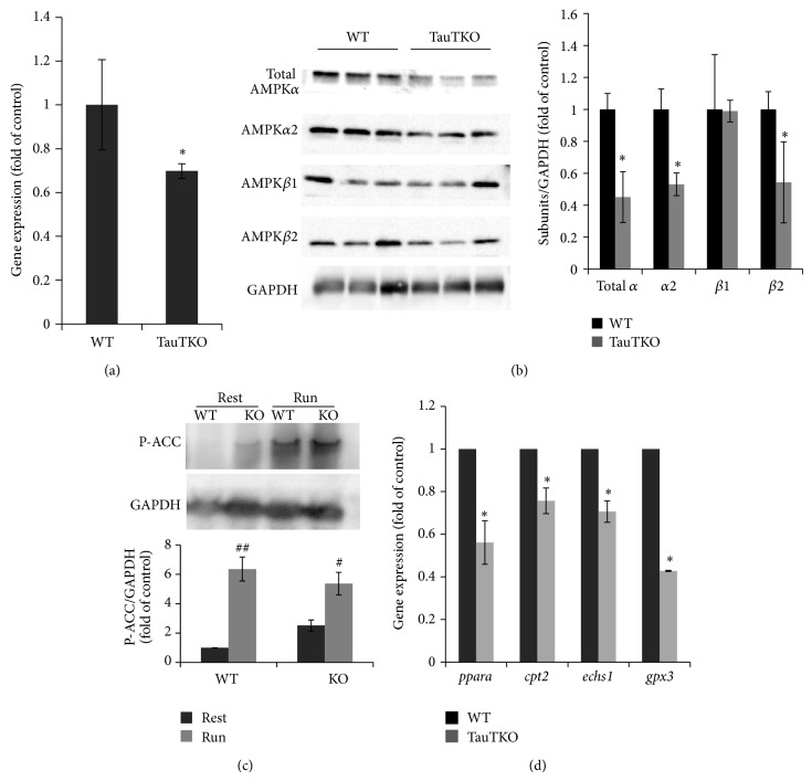
Figure 3.
Expression of AMPK subunits and genes of PPARα and its targets in tibial anterior muscle of WT and TauTKO mice. (a) Gene expression of AMPK β2 was measured by qRT-PCR method. n = 4. * P < 0.05 versus WT. (b) AMPK subunits (α1, α2, β1, and β2) and GAPDH in TA muscle of WT and TauTKO mice were detected by Western blot. n = 3. * P < 0.05 versus WT. (c) Phospho-ACC (Ser79) and GAPDH in skeletal muscle before and 20 min after treadmill running were detected. Data are mean ± se. n = 3–6. # P < 0.05, ## P < 0.01 versus rest group. (d) The change in genes of PPARα (ppara), carnitine palmitoyl transferase 2 (cpt2), short-chain enoyl CoA hydratase (echs1), and glutathione peroxidase c (gpx3) was analyzed by quantitative RT-PCR. Data are mean ± SE. n = 4–7. * P < 0.05 versus WT.

