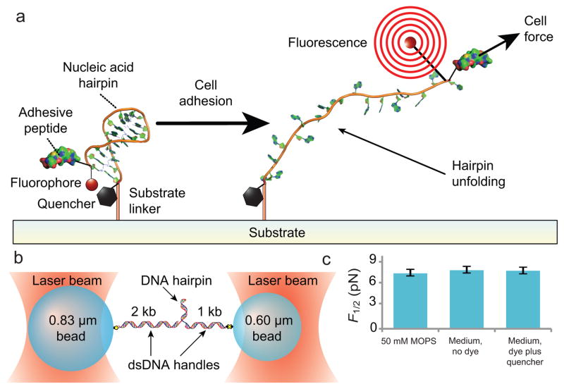
Figure 1. Design and characterization of DNA hairpin force probe.
(a) Schematic depiction of the TPs. A DNA hairpin is functionalized with a fluorophore-quencher pair, covalently conjugated by its 3′ end to a solid substrate, and conjugated at its the 5′ end, via a PEG spacer, to the integrin-binding peptide RGD. Upon the application of sufficient force to unfold the hairpin, the fluorophore separates from the quencher and fluoresces. (b) Schematic of the experimental geometry used to characterize the mechanics of the hairpins. The DNA hairpin is attached at each of it ends to dsDNA handles bound to optically trapped beads (not to scale) in a force-clamped arrangement. (c) Measured F1/2 (hairpin opening force) values as a function of media and fluorophore-quencher conjugation at 21 °C (mean ± s.e.m.). n>3 for each condition.

