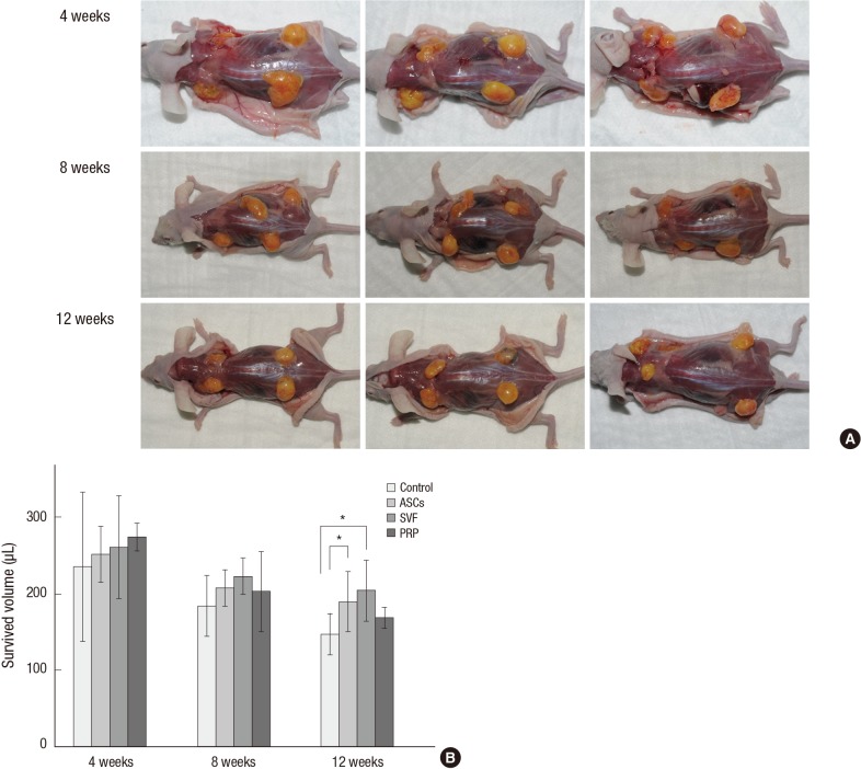
Fig. 2.
Surviving volume of grafted human fat tissue. (A) The macroscopic appearance showed that neovasculature structures were observed in the adjuvant groups after 8 weeks. (B) Volumes of the surviving adipose tissues. For the ASCs and SVF groups, the mean volumes were significantly greater than that of the control at 12 weeks. Mean and 95% CI are shown (*P < 0.05). ASCs, adipose-derived stem cells; SVF, stromal vascular fraction; PRP, platelet-rich plasma.

