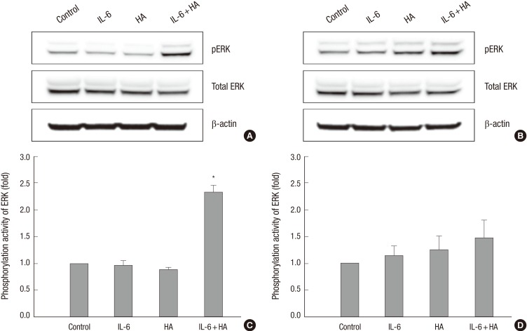
Fig. 3.
Phosphorylation of ERK by treatments of IL-6 and HA in HaCaT cells. (A) HaCaT cells were treated with IL-6 and HA for 1 hr. Phosphorylation of ERK was significantly increased by combining IL-6 and HA treated group. (B) HaCaT cell lines were treated with IL-6 and HA for 24 hr. Phosphorylated ERK was no significant difference for 24 hr all treated groups. Phosphorylation and total protein expression of ERK were detected by Western blot analysis. Densitometry analysis of ERK phosphorylation is showed as fold change versus control at 1 hr (C) and 24 hr (D). Data are presented as the means±SEM of three experiments. *P = 0.009 compared to the control group.

