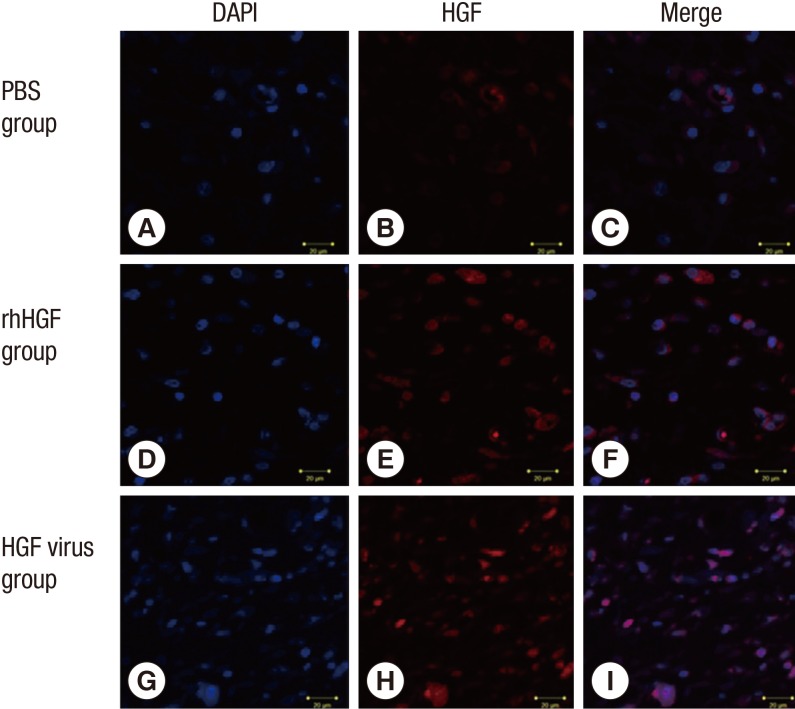
Fig. 2.
Immunofluorescence staining of HGF 10 days after flap elevation. Sections (3 µm thick) of rat skin flap tissue (distal part) were stained with DAPI to label nuclei (A, D, G) and antibodies against HGF (B, E, H). Scale bars (50 µm) are indicated in all photomicrographs. Merged immunofluorescence images reveal that HGF is expressed in and around DAPI-stained cells (C, F, I).

