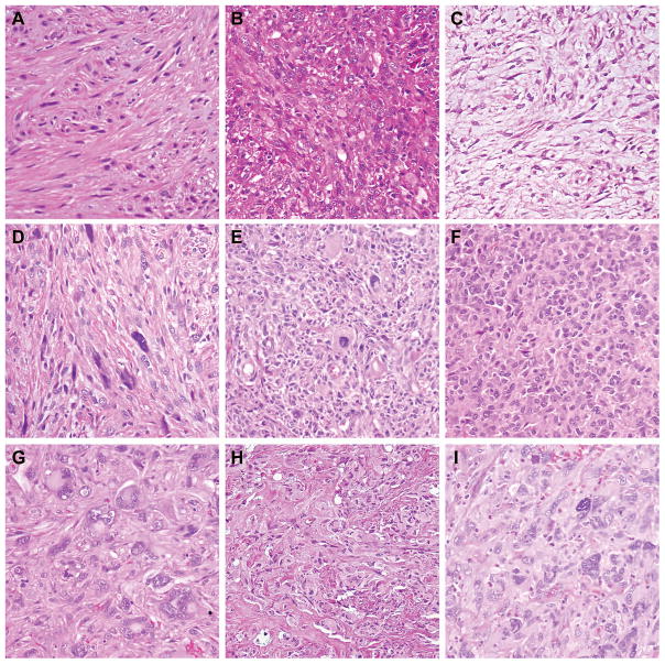
Figure 1. Morphologic Variation in Leiomyosarcoma.
A) Well differentiated tumor demonstrating intersecting fascicles of slightly atypical eosinophilic spindle cells. B) Moderately differentiated tumor with nuclear variability and increased disorganization of fascicles. C) Moderately differentiated myxoid tumor with bland spindle cells in a storiform to fascicular arrangement. D) Moderately differentiated tumor with scattered “monster cells.” E) Poorly differentiated tumor with loss of fascicular architecture, and increased rounded to epithelioid cells. F) Poorly differentiated tumor with epithelioid features. G) Poorly differentiated tumor showing therapy effect and marked nuclear pleomorphism. H) Poorly differentiated tumor with pleomorphism, loss of architecture, and numerous mitoses. I) Poorly differentiated “undifferentiated pleomorphic sarcoma-like” tumor showing storiform growth, marked pleomorphism and scattered inflammatory infiltrate. (All panels at 200X).

