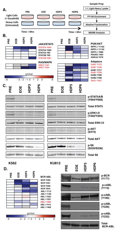
Figure 1. Transient Exposure of CML Cell Lines to Dasatinib Results in Durable Dephosphorylation of Select Tyrosine Residues in Myeloid Growth-Factor Receptor Signaling Pathways.
A. Schematic of SILAC-based quantitative phosphoproteomic analysis of global phosphotyrosine signaling in the K562 cells before and after a high-dose pulse (HDP) of dasatinib. K562 cells grown in “light” (non-isotope-containing) RPMI were treated with a 100nM dasatinib for 20 minutes, and cell lysates were generated before HDP (PRE), at the time of drug washout (EOE), and 3hr and 6hrs post-HDP (HDP3, HDP6). Equivalent lysates were generated from K562 cells grown in “heavy” (isotope-containing) RPMI. Light and heavy K562 cell lysates were mixed at a 1:1 ratio prior to phosphotyrosine peptide (PY100) enrichment, peptide fractionation, and MS/MS analysis.
B. Heat map representation of persistent phosphorylation changes in myeloid growth factor receptor signaling pathways identified by bioinformatic functional analysis. Change in phosphorylation at each HDP time point was normalized to the “PRE” condition and are represented on a log2 - transformed scale. Gray areas designate “no data”.
C. Western immunoblot analysis of select myeloid growth factor receptor signaling pathways in K562 and KU812 cells before and after a 100nM HDP of dasatinib.
D. Heat map representation of BCR-ABL phosphorylation identified by phosphoproteomic analysis and western immunoblot analysis in K562 cells before and after a 100nM HDP of dasatinib. (ABL1a numbering).

