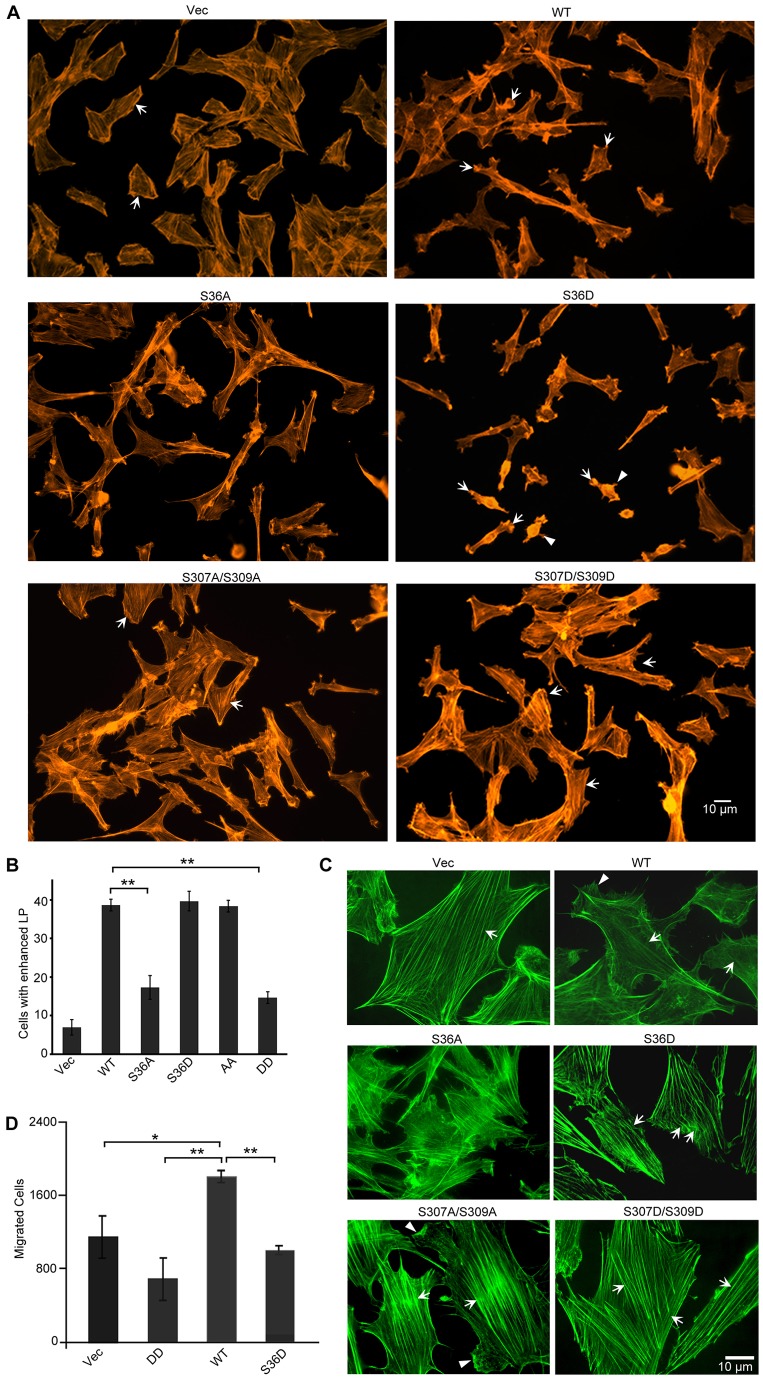
Fig. 2.
Actin cytoskeletal alterations caused by expression of CAP1 phospho mutants. (A) Wide-field fluorescence imaging shows alterations in lamellipodia and stress fibers in cells that express the mutants. The actin cytoskeleton was stained with fluorescently tagged phalloidin (Alexa-Fluor 594). Cells indicated by arrows show the typical changes to the actin cytoskeleton, as discussed for S36D- and S307/S309-mutant-expressing cells; arrowheads indicate randomly positioned protrusions in the S36D-expressing cells. (B) Percentage of cells with prominent lamellipodia. Fifty cells were counted for each cell type and the experiment was repeated for three times. The data were analyzed using Student's t-test and shown as mean ± s.d; **P <0.01). (C) Confocal fluorescence microscopy showing further details of the actin cytoskeletal alterations. F-actin was stained with fluorescently tagged phalloidin (Alexa-Fluor 488) and images were taken using a 60×lenses with a BD Pathway 855 imaging system (GFP is stably expressed at low levels and does not interfere with Alexa-Flour 488 in microscopy). Arrows indicate changes to the stress fibers of each cell type, as discussed; arrowheads indicate lamellipodia. (D) Transwell migration assays revealed reduced motility in both S36D and DD cells. The data were analyzed and are shown as in B; *P<0.05, ** P<0.01. The experiment was repeated three times with similar results.

