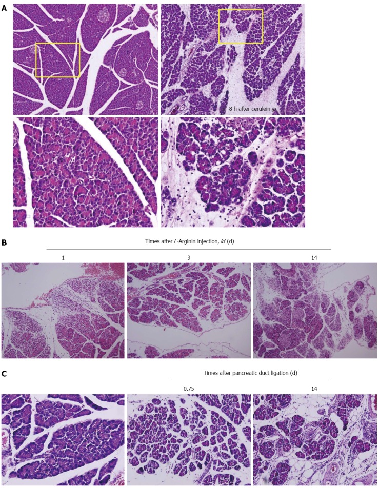
Figure 1.

Animal models for pancreatitis. A: Cerulein-induced edematous pancreatitis. Caerulein-induced pancreatitis is a valuable experimental model for studying altered intracellular transport, compartmentation of lysosomal, and digestive enzymes, resulting in edematous pancreatitis. The formation of enlarged secretory vacuoles containing lysosomal and digestive enzymes is paralleled by the activation of lysosomes and degradation of cellular organelles in autophagosomes. On the level of secretory and autophagic vacuoles, activation of serine proteases occurs, which in addition to increasing lysosomal enzyme activities can represent the initial stage for acinar cell destruction and the development of pancreatitis; B: L-arginine-induced necrotizing pancreatitis. Parenchymal hemorrhage and widespread acinar cell necrotic changes were noted with L-arginine; C: Pancreatic duct ligation-induced pancreatitis. Morphologic examination of the pancreas showed massive interstitial edema, apoptosis, and necrosis of acinar cells with infiltration of neutrophil granulocytes and monocytes 0.75 d after pancreatic duct ligation. Two weeks later after periodontal ligament, the destructed parenchyma with fat replacement as well as some fibrotic changes were seen.
