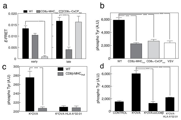
Figure 3. Two stage interaction between TCR-CD8.
(a) OT-I hybridomas expressing CD8αWT, CD3ζ-eGFP, and CD8β-mCherry (black); CD8αWT, CD3ζ-eGFP, and CD8β-MHCmut-mCherry (dark grey) or CD8α-CxCPmut, CD3ζ-eGFP, and CD8β-mCherry (light grey) were added to lipid bilayers containing Kb- OVA and ICAM-1, fixed at 1 min (“early”) or 10 min (“late”), and imaged by TIRFM at the immune synapses. FRET efficiency ±s.e.m. was plotted, “early” WT (n=30), CxCPmut (n=25), MHCmut (n=28), and “late” n=28, 24, 29, respectively. (b) OT-I hybridomas expressing CD8αβ WT (black); CD8αβ-MHCmut (dark gray) or CD8α-CxCPmut and CD8β WT (light gray) were stimulated as in (a) for 15 min and stained with anti- phospho-Tyr. Images were taken using TIRFM. Mean fluorescence of WT n=20, MHCmut n=33, CxCPmut n=36 and VSV n=21 ±s.e.m. is shown. OT-I cells expressing CD8αβ-WT (white) were added to bilayers containing Kb-VSV and ICAM-1 as a negative control. (c) OT-I hybridomas expressing CD8αβ-WT (black) or CD8αβ-MHCmut (dark gray) were added to bilayers containing either Kb-OVA or Kb-OVA-HLA-A*02:01 plus ICAM-1 for 15 min, then stained with anti-phospho-Tyr. Mean fluorescence ±s.e.m. shown for Kb- OVA, WT n=15; CD8β-MHCmut n=15 and for Kb-OVA-HLA-A*02:01 WT n=15; CD8β-MHCmut n=19. (d) Primary mouse OT-I CTL were pretreated with blocking anti-CD8 (dark gray) or directly (black) added to bilayers containing either Kb-OVA or Kb-OVA- HLA-A*02:01 plus ICAM-1 or ICAM-1 alone (control) for 15 min, then stained with anti-phospho-Tyr. Mean fluorescence of control n=23, Kb-OVA n=17, anti-CD8β n=19 and Kb-OVA-HLA-A*02:01 n=20 ±s.e.m. are shown. Unpaired t-test. ***p<0.0001.

