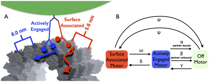
Figure 2. Experimental and Model Arrangements.
(a) Our experimental setup consists of a 440-nm diameter polystyrene bead as the cargo (green), with two recombinant K560 half-height kinesins (blue, AE kinesin; red, SA kinesin) connected through C-terminal histidine tags to a single monoclonal antibody (dark blue). Below the motors is shown a 13-protofilament right-handed A-lattice microtubule39. The microtubule kinesin binding domain lattice is depicted; dark grey tubulins actively bind kinesin heads, while light grey tubulins interact through non-specific interactions. The transverse distance of 5.6 nm between microtubule protofilaments appears in red, and the axial spacing of 8 nm between kinesin binding locations appears in blue. (b) Transition rate variables and states in our dynamic two-motor state transition model.

