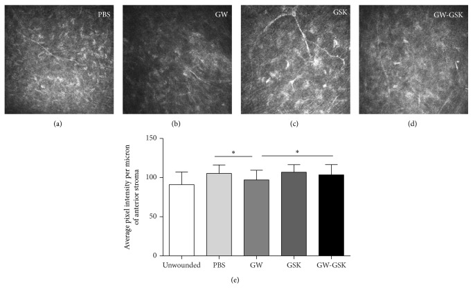
Figure 4.
In vivo confocal micrographs of the anterior stroma of similar depth four weeks ((a)–(d)) after laser ablation. Corneas in the GW501516 group (b) showed quiescent keratocytes with low reflectivity, while the corneas in the GSK3787 group (c) showed active keratocytes with high reflectivity. (e) The relative intensities of reflectivity were evaluated based on the average pixel intensity per micron of the central anterior stroma. The data are presented as the mean ± SD (n = 4–6), and differences were analyzed by ANOVA. * P < 0.05, ** P < 0.01 versus GW501516 group.

