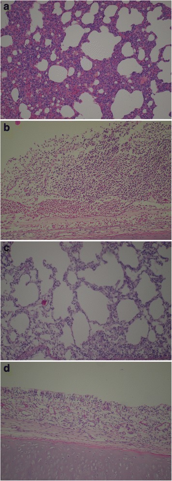
Figure 3.

Representative photomicrographs of (a, c) lung tissue and (b , d) nasal mucosa from rabbits infected intranasally with P. multocida. (a) Lung from the positive control (untreated) animals showing interstitial inflammatory reaction, and (c) normal lung from the β-glucan treated animals. (b) Nasal mucosa from the positive control (untreated) animals showing epithelial necrosis and intensive heterophil granulocyte infiltration with submucosal edema. (d) Normal nasal mucosa from the β-glucan treated animals. Hematoxylin and eosin stain; magnification, 100×.
