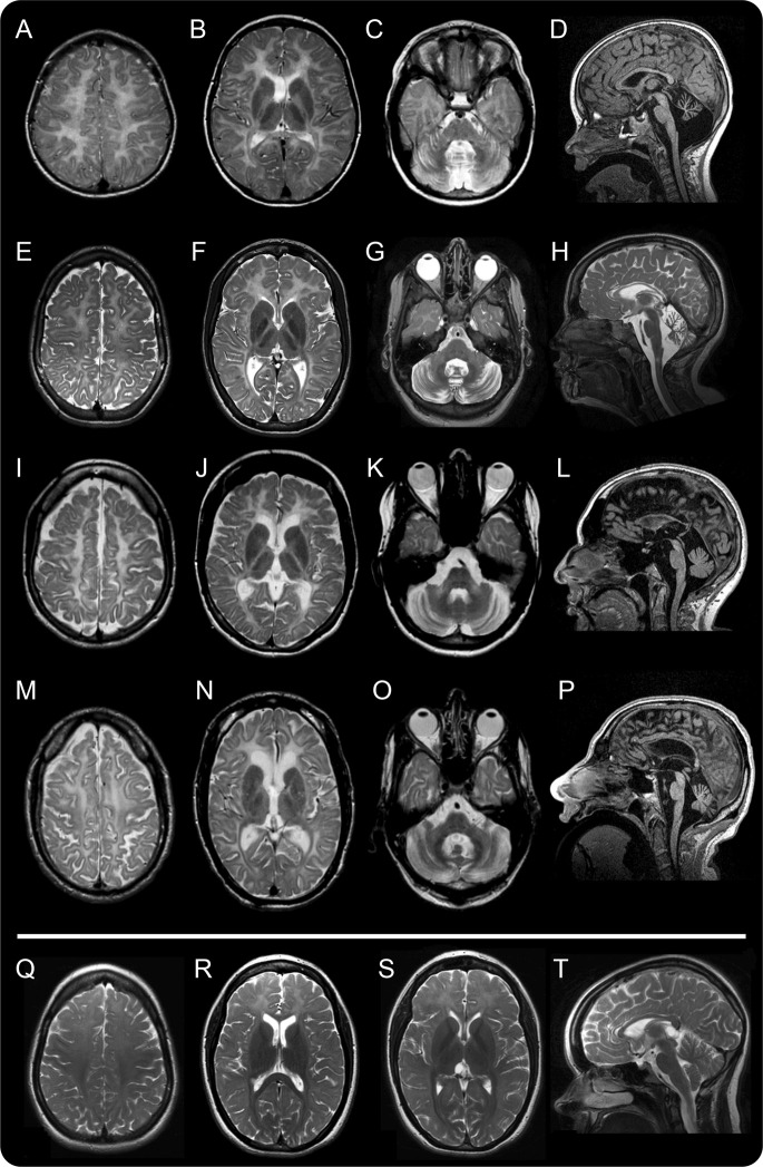
Figure 2. MRI changes with age.
MRI of 4 patients with 4H syndrome at 6 (A–D, patient 4H-52, row 1), 12 (E–H, patient 4H-53, row 2), 23 (I–L, patient 4H-58, row 3), and 40 (M–P, patient 4H-94, row 4) years of age. The axial T2-weighted images all show diffusely elevated white matter signal, less hyperintense than CSF, consistent with hypomyelination. The signal of the optic radiations is hypointense, indicating myelination. In the youngest patient, there is a small hypointense dot in the posterior limb of the internal capsule (B). All patients show a relatively hypointense signal of the ventrolateral thalamus. The corpus callosum becomes thinner with age (sagittal T1 images, A, L, P, and sagittal T2 image, H). The sagittal images also demonstrate cerebellar atrophy. White matter signal of the middle cerebellar peduncles and the cerebellar white matter is too high on the T2-weighted images; the dentate nucleus appears hypointense as well as the dorsal tegmentum (C, G, K, O). (Q–T) MRI of patient 4H-45 homozygous for c.1586T>A (POLR3B) at age 16 years. Myelination of the perirolandic cortex and the parieto-occipital white matter is relatively good on these axial T2-weighted images, compared to the frontal white matter (Q, R, S). The splenium and the anterior limb of the internal capsule (R) as well as the optic radiations (S) are well-myelinated. There is a small lesion in the right optic radiation (R). No cerebellar atrophy is seen on the sagittal T1-weighted image (T).

