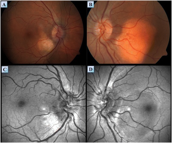
Figure 7.

Fundoscopy of patient #1. Fundoscopy images of the right (A) and left (B) eyes of patient #1, demonstrating sub choroidal lesions involving the macula densa. (C,D) Red free imaging produced by a blue wavelength confocal scanning laser ophthalmoscope demonstrating the extent of these lesions in the right (C) and left (D) eyes.
