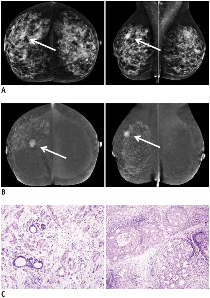
Fig. 2.

Infiltrating ductal carcinoma (arrows) of right breast in 63-year-old woman with dense fibroglandular breast tissue.
A. Digital conventional mammography (MG) with mediolateral oblique (MLO) view of right breast reveals oval, well demarcated shadow 15 mm in diameter. This mass is not visible in MG craniocaudal (CC) projection. B. Contrast-enhanced spectral mammography in same patient shows poorly demarcated focus of enhancement 15 mm in diameter visible in both MLO and CC projections. Additionally, in right breast, small enhancing foci are visible in upper and lower outer quadrants. C. In histopathological examination, infiltrating carcinoma (of no special type) grade 2 was found within main focus of enhancement. Ductal carcinoma in situ (DCIS) component was noticed in unaltered areas macroscopically. Hematoxylin and eosin staining. Objective magnification 10 × (invasive component, left) and 20 × (DCIS, right).
