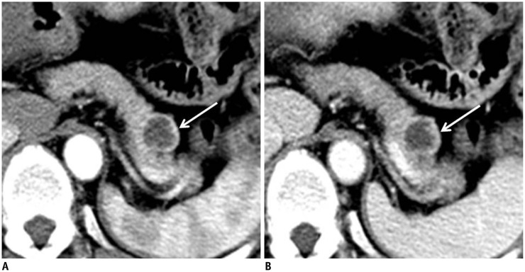
Fig. 2.
Pancreatic neuroendocrine tumor in 54-year-old woman.
A, B. Transverse contrast-enhanced arterial (A) and portal (B) phase computed tomography images demonstrate 2-cm round cyst-like mass in pancreatic tail. Thick (3.3 mm) uneven wall (white arrows) shows high attenuation in arterial phase and iso-attenuation in portal phase.

