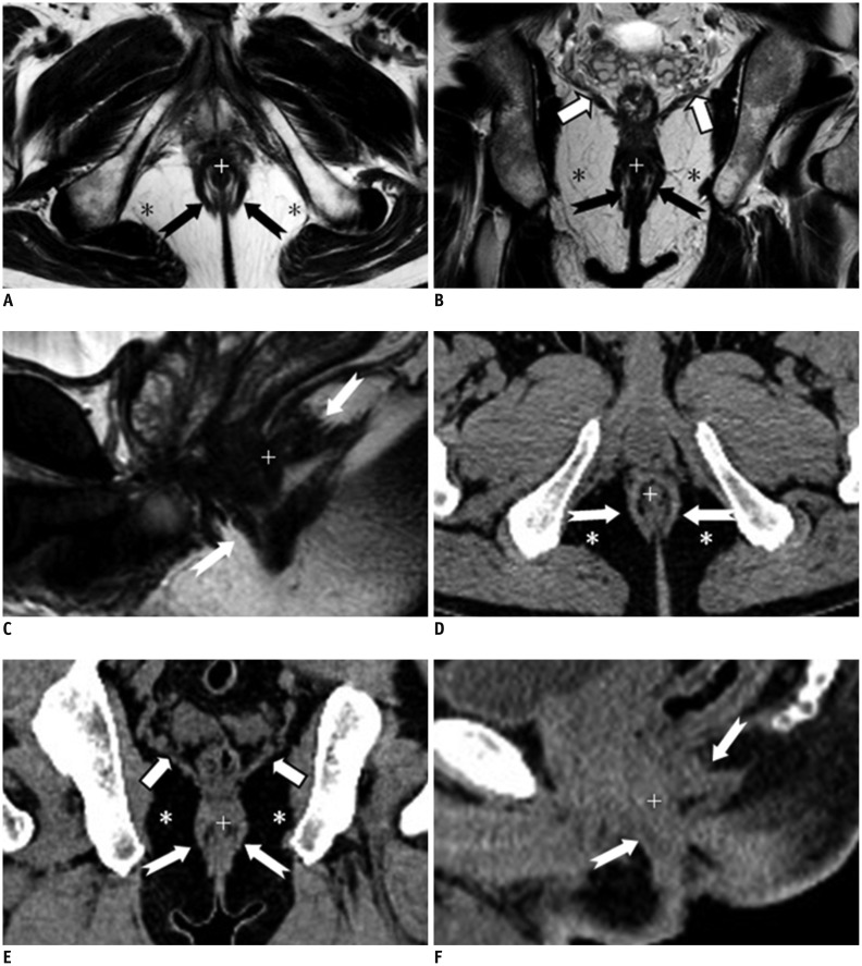
Fig. 2.
Normal perianal anatomy of 45-year-old male volunteer imaged with CT and MR imaging.
MR imaging (T2-weighted without fat suppression images; A-C) and CT (D-F): anal sphincter complex is seen as two concentric rings. Inner internal sphincter (+), outer external sphincter (white or black dovetail arrows), levator ani muscle (thick arrows), fat containing ischioanal fossa (*).

