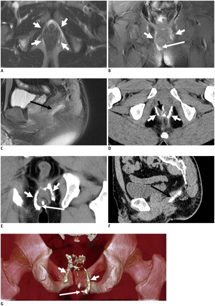
Fig. 8.
38-year-old male with semi-horseshoe fistula.
CT fistulography (D-G) and MR imaging (T2-weighted with fat suppression in A-C): transverse, coronal images clearly show circumferential spread of fistula (short white arrows in A-G). Extent of disease and complicated spatial information are better seen on volume rendering image (short arrows in G). External opening (long white arrow in B, E, G), internal opening (short black arrow in D), and secondary ramification (long black arrow in C, F) are seen.

