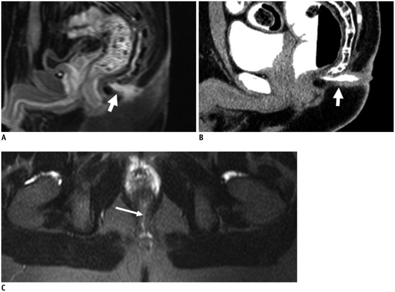
Fig. 9.
26-year-old male with fistula.
MR imaging (T1-weighted with fat suppression in A, T2-weighted with fat suppression in C) and CT fistulography (B): fistula (short white arrow in A, B) spreads backward to skin's surface with evident external opening. Tenuous internal opening (long white arrow in C) was successfully identified on MR image. Internal opening is confirmed clearly on MR image but not corresponding CT image, owing to lack of contrast agent filling.

