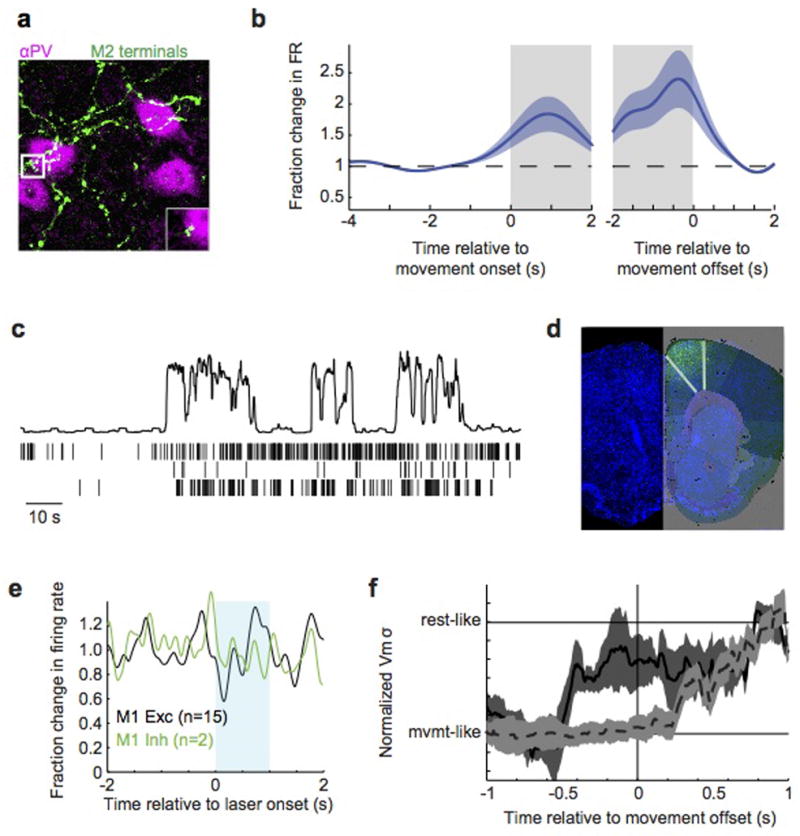
Extended Data Figure 8. M2 activity drives motor-related changes in auditory cortical dynamics.

a, Z-stack micrograph of M2 axons (green, AAV-GFP injection) forming appositions with PV+ immunostained interneurons in auditory cortex (magenta). Inset shows a high magnification single (2um) optical section of an apposition. b, M2 spiking activity relative to movement onset (left) and offset (right), normalized to pre-movement activity. c, Three simultaneously recorded M2 neurons during three transitions from rest to movement. Top panel shows movement extracted from video (a.u.). d, Cell bodies and local terminal field of ChR2+ neurons following injection of AAV.2/1.ChR2 into M2. Image is overlaid with an atlas from the Allen Brain Institute. e, Extracellular recordings in M1 of VGAT-ChR2 mouse during blue laser stimulation over ipsilateral M2 showing no change in firing of neurons with broad (black, putative excitatory) or narrow (green, putative inhibitory) neurons. f, Change in membrane potential variance of auditory cortical excitatory neurons over time during optogenetic silencing of either ipsilateral (black, solid) or contralateral (gray, dashed) M2. For each neuron, the time-varying membrane potential variance was measured during a sliding window that extended 500 ms into the past. Traces were then averaged across neurons after aligning each to the time of movement cessation. Silencing ipsilateral M2 causes membrane potential variance to change prior to movement offset, whereas silencing contralateral M2 causes the variance to change after movement offset.
