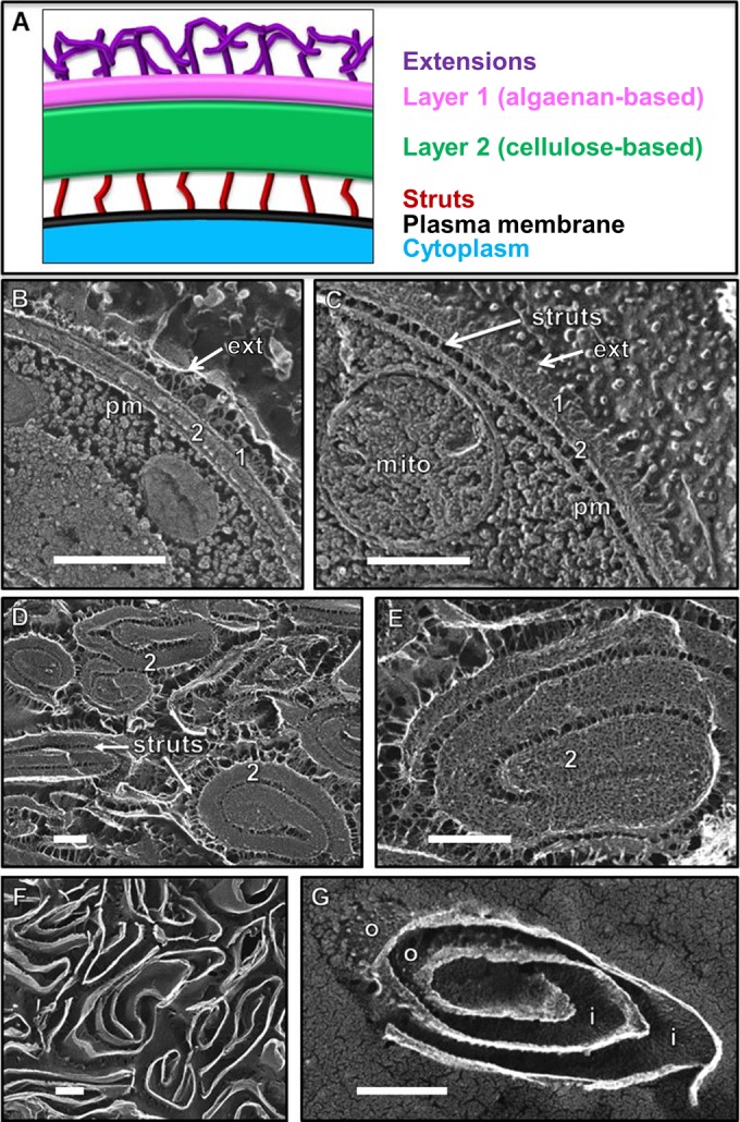
FIG 2.

(A) Model of the Nannochloropsis wall based on quick-freeze deep-etch EM (QFDE-EM) images. The native layer 1 adopts a TLS configuration in thin section (2) but forms a single layer in replicas and is overlain with fibrous extensions. Layer 2 is thicker and often associates with the plasma membrane via narrow struts. (B) Cross fracture through the native wall showing its two layers (1, 2), distal extensions (ext), and close association with the plasma membrane (pm). (C) Tangential fracture through the two native wall layers showing distal extensions (ext) and proximal struts attaching to the plasma membrane (pm). mito, mitochondrion. (D and E) Pressed cell walls, with the prominent fibrillar layer 2 masking the presence of layer 1. (F and G) Same pressed wall preparation as shown in panels D and E after 24 h of incubation in cellulase. Fibrillar layer 2 and struts were digested, exposing layer 1, whose outer (o) and inner (i) faces are granular. All scale bars, 250 nm.
