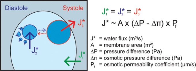
FIG 1.

Osmoregulation in Chlamydomonas reinhardtii. Water fluxes across plasma and CV membranes are indicated. The CV on the left is representative of early diastole. At the end of diastole, the large CV (right CV; approximately 2 μm in diameter) develops from small (diameter, 100 to 200 nm) vacuoles (broken blue arrow). The CV on the right depicts the transition from diastole to systole phase. The contractile vacuole associates closely with the PM and expels liquid into the medium. During this process, the CV collapses and fragments into numerous small vacuoles (broken red arrow). To sustain cell size, water fluxes during the three water transport steps must be in equilibrium (J1* = J2* = J3*). The comparison of the CV volume at the end of diastole to the total volume of small vesicles (same amount of membrane material; diameter, 100 to 200 nm) at the beginning of diastole indicates that at least 90% to 95% of the CV volume is discharged during a single CV cycle (assuming ideal spheres for vesicles and the large CV). As the collapsed membrane material of the CV rarely forms ideal spheres, the real percentage of discharge is even higher. For simplicity, the CV volume at the end of diastole is used in all calculations, which gives rise to a small overestimation (less than 5%) of water flux and the calculated membrane permeability coefficient, Pf.
