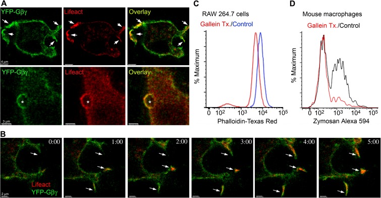
FIG 5.
Involvement of Gβγ subunits in macrophage phagocytosis. (A) Confocal microscopy images of RAW 264.7 cells expressing RFP-Lifeact (red), ½ YFP-Gβ1, and ½ YFP-Gγ2 exposed (bottom) or not (top) to zymosan. Sites of strong overlap are indicated with arrows (top). Bar, 4 μm. Location of zymosan is indicated with an asterisk (bottom). Bar, 3 μm. (B) Time-lapse confocal microscopy of RAW 264.7 cells expressing RFP-Lifeact (red), ½ YFP-Gβ1, and ½ YFP-Gγ2. Arrows show cell protrusions where YFP-Gβ1γ2 and RFP-Lifeact eventually colocalized. Bar, 2 μm. (C) Flow cytometry of RAW 264.7 cells treated with gallein, or not, and stained for phalloidin. RAW 264.7 cells were treated overnight with 10 μM gallein prior to phalloidin staining and flow cytometry. (D) Flow cytometry of mouse BMDM treated with gallein (20 μM, 15 min) or not and then exposed to fluorescent zymosan (10 μg) for an additional 30 min.

