FIG 1.
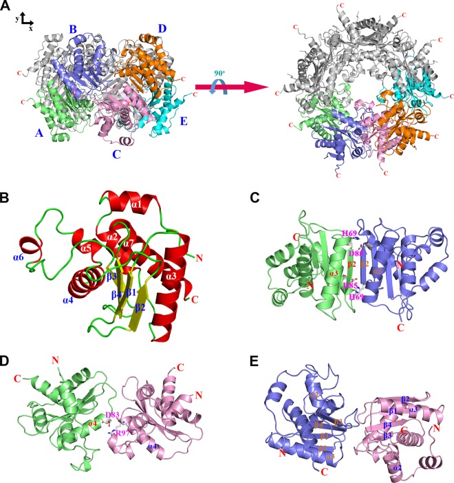
Structural features of Ybr137wp. (A) Decamer conformation of Ybr137wp. The five Ybr137wp molecules in an asymmetric unit are designated Mol A to Mol E and are shown in green, blue, pink, orange, and cyan, respectively. The left and right panels are the side view and top view of the decamer, respectively. (B) Ribbon diagram of the Ybr137wp monomer. The four β-strands (β1 to β4), seven α-helices (α1 to α7), and loops in the protein are shown in yellow, red, and green, respectively. (C to E) Three types of interface interactions between subunits, the AB-type interface (C), the AC-type interface (D), and the BC-type interface (E). The corresponding molecules are colored as described above for panel A. The residues involved in salt bridge formation are depicted as a stick model.

