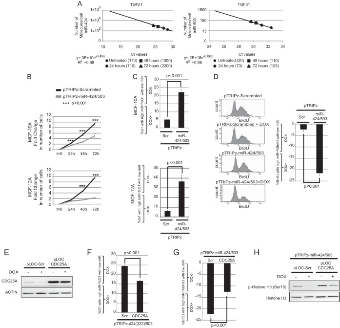
FIG 4.
Increased miR-424/503 expression levels impair the G1/S transition and compromise cell proliferation. (A) Estimation of the numbers of molecules of miR-424 and miR-503 per cell during TGF-β exposure at several time points in MCF-10A cells as described in the main text. (B to D) MCF-10A and MCF-12A cells were lentivirally transduced with a control (pTRIPz-Scrambled) or with a Dox-inducible vector expressing miR-424(322)/503 (pTRIPz-miR-424/503). The cells were treated for 3 days with 100 ng/ml of doxycycline (+Dox) or left untreated for the same time (−Dox). Cell proliferation was then monitored for 72 extra hours using the CellTiter-Glo system. The results indicate that the doxycycline-induced miR-424/503 impairs cell proliferation due to accumulation of cells in G1 phase (C) and a reduction of BrdU-positive cells (D). The y axis in cell cycle graphics represents the percentage of cells in G1 when levels of the miRNA cluster are high (DOX+) minus the percentage of cells in G1 when levels of miRNA cluster are low (DOX−). The same is applicable for the graphics representing BrdU incorporation data. (E) Analysis by Western blotting of pT-miR-424/503-MCF-10A cells transduced with lentiviral particles containing a control vector (pLOC-Scr) or nontargetable pLOC-CDC25A devoid of the 3′ UTR. (F and G) Expression of nontargetable CDC25A in Dox-treated pT-424/503-MCF-10A cells attenuates the G1 arrest imposed by miR-424/503 (F) and the decrease in BrdU-positive cells (G). (H) Representative Western blot showing that the expression of the nontargetable CDC25A form ensures progression into mitosis in Dox-treated pT-424/503-MCF-10A cells as measured by p-histone 3.

