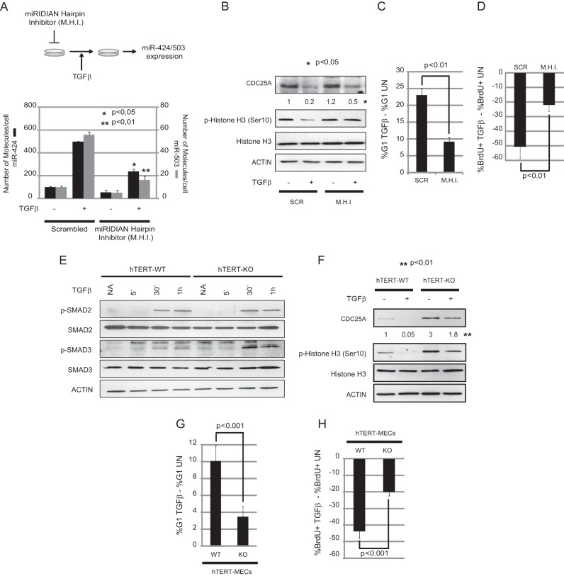
FIG 6.
MiR-424/503 inhibition attenuates TGF-β cytostatic effects. (A) MCF-10A cells were transfected with miRIDIAN Scrambled (Scr) or hairpin inhibitors (M.H.I.) for miR-424(322) and miR-503 and treated with TGF-β-1 ligand for 24 h (A, upper panel). Transfection of M.H.I. results in reduction of the levels of both miRNAs as measured by qRT-PCR analysis (A, bottom panel). (B) Analysis by Western blotting shows that M.H.I.-transfected cells are able to rescue the decrease in CDC25A protein levels after TGF-β treatment. Numeric values show densitometry quantification of the bands. Western blot analysis also shows reentry into mitosis as measured by p-histone 3. (C and D) Mir-424(322) and miR-503 knockdown cells show attenuated arrest in G1 phase (C) and a decrease in BrdU-positive cells (D) imposed by TGF-β. (E) Western blot analysis shows that both wild-type and miR-424−/−/miR-503−/− hTERT-MECs respond to TGF-β by activating p-SMAD2 and p-SMAD3 at similar levels. (F) Analysis by Western blotting shows that miR-424−/−/miR-503−/− hTERT-MECs are not able to downregulate CDC25A protein levels in response to TGF-β to the levels observed in their wild-type counterparts. Immunostaining for p-histone 3 indicates that the transition into mitosis is compromised in wild-type hTERT-MECs but not in miR-424−/−/miR-503−/− cells after TGF-β exposure. (G and H) Analysis of the percentage of cells in G1 phase (G) and BrdU incorporation ratios (H) indicates that miR-424−/−/miR-503−/− hTERT-MECs overcome growth-inhibitory effects induced by TGF-β exposure. In panels C, D, G, and H, the data are presented as the ratio between results for TGF-β-treated cells (TGFβ) and those for untreated cells (UN). All the experiments were performed in triplicate.

