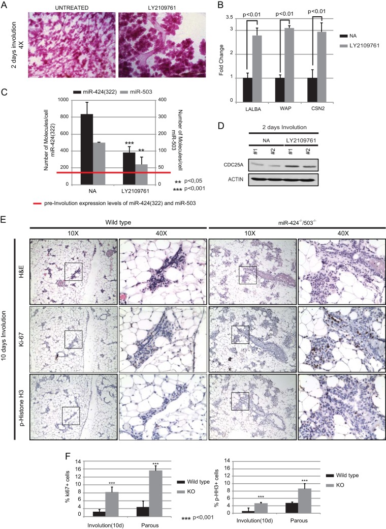
FIG 7.
Inhibition of TGF-β signaling during mammary involution abrogates the induction of miR-424 and -503 expression. (A and B) Administration of the TβRI/II specific inhibitor LY2109761 partially inhibits mammary epithelial regression as measured by whole-mount carmine red staining (A) and quantification by qRT-PCR of the milk proteins α-lactalbumin (LALBA), whey-acidic protein (WAP), and β-casein (CSN2) in purified mammary epithelial cells (B). (C) Pharmacological inhibition of the TGF-β pathway abrogated the induction of miR-424 and -503 expression levels. The red line indicates the number of molecules observed for miR-424 and -503 during lactation. (D) Western blot showing increased protein levels of CDC25A in mammary epithelial cells treated with LY2109761. (E) Immunohistochemistry studies performed with mammary glands at 10 days of involution by using hematoxylin and eosin (H&E), Ki67, and p-histone 3 in wild-type and miR-424−/−/miR-503−/− females. These studies show a nearly complete regression in wild-type glands, while miR-424−/−/miR-503−/− females exhibit a delayed involuting phenotype coupled with an increased proliferative activity, as shown by increase in Ki67- and p-histone 3-positive cells. (F) Quantification of percentages of Ki67- and p-histone 3-positive cells in mammary glands at 10 days and 60 days postinvolution. All the experiments were performed in triplicate. A minimum of three animals were analyzed. For quantification of percentages of Ki67- and p-histone 3-positive cells, a minimum of 3,000 cells were counted per animal.

