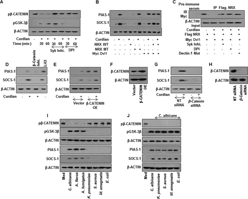
FIG 6.
SyK-ROS-stabilized β-catenin mediates dectin-1 ligand-induced expression of PIAS-1 and SOCS-1. (A) Immunoblotting analysis of pβ-catenin and pGSK-3β in whole-cell lysates of mouse macrophages pretreated with Syk inhibitor or DPI for 60 min followed by treatment with curdlan. (B) Expression analysis of PIAS-1 and SOCS-1 by immunoblotting in RAW 264.7 macrophages transiently transfected with the indicated OE constructs followed by treatment with curdlan for 12 h. (C) Immunopulldown (IP) with anti-Flag antibody followed by immunoblot analysis for Myc in RAW 264.7 macrophages transiently transfected with respective constructs in the presence or absence of SyK inhibitor or DPI. (D) Mouse peritoneal macrophages were pretreated with β-catenin inhibitor for 60 min or with LiCl alone for 4 h and then treated with curdlan for 12 h. Expression of PIAS-1 and SOCS-1 was analyzed by immunoblotting. (E) RAW 264.7 macrophages were transiently transfected with β-catenin OE followed by treatment with curdlan. Total cell lysates were analyzed for PIAS-1 and SOCS-1 by immunoblotting. (F) Validation of β-catenin OE construct. (G and H) Peritoneal macrophages from C3H/HeJ mice were transiently transfected with β-catenin siRNA followed by treatment with curdlan for 12 h. Lysates were analyzed for PIAS-1 and SOCS-1 expression (G) and β-catenin levels (H) by immunoblotting. (I and J) Mouse peritoneal macrophages from C3H/HeJ mice were infected with the indicated fungi and bacteria (I) or coinfected with C. albicans followed by the tested bacteria (J) for 60 min to assess pβ-catenin and pGSK-3β levels and for 12 h to assess PIAS-1 and SOCS-1. All blots are representative of 3 independent experiments. WT, wild type; MT, mutant; NT, nontargeting; IB, immunoblot; OE, overexpression construct.

