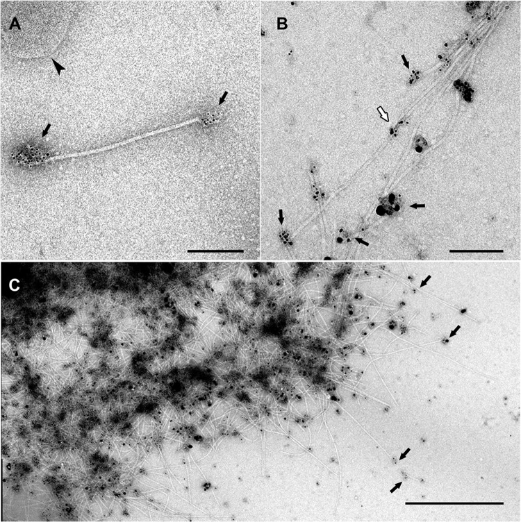
FIG 1.
Immunogold labeling of Iho670 fibers in solution. The labeling of purified fiber proteins was performed using the rabbit anti-Iho670 primary antibody (dilution of 1:10) and the goat anti-rabbit secondary antibody (6-nm gold, dilution of 1:5 [A]; ultrasmall gold, dilution of 1:5 [B]) with subsequent silver enhancement for 6 min. The images show single filaments (A) or aggregates of filaments (B, C). In most cases, the antibodies bind to the ends of the filaments where epitopes can be accessed (black arrows). Binding to intermediate regions is rare (white arrow). Sometimes, small pieces of membranes can be found after the purification (arrowhead in panel A). Scale bars, 200 nm (A and B) and 1 μm (C).

