FIG 2.
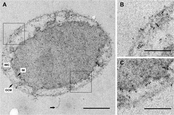
Immunogold localization of Iho670 on ultrathin sections. Labeling was performed on sections of high-pressure frozen, freeze-substituted, and epon-embedded I. hospitalis cells. (A) Ultrathin section of an I. hospitalis cell with a fiber, indicated by black arrows. Primary antibody, rabbit anti-Iho670 (dilution of 1:100); secondary antibody, goat anti-rabbit antibody and 6-nm gold (dilution of 1:50). White arrows indicate the labeling of tubes/vesicles in the intermembrane compartment. At higher magnification, the selected areas in panel A exhibit signals in the outer cellular membrane (B) and the inner membrane (C). Scale bars, 500 nm (A) and 200 nm (B, C).

