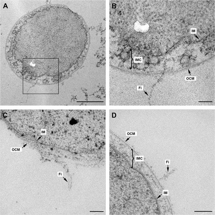
FIG 3.
Illustration of I. hospitalis fibers in ultrathin sections. (A to D) Ultrathin sections of I. hospitalis cells exhibiting longitudinal cuts, including fibers. (B) At higher magnification of the selected area in panel A, the connection of the fiber with the cell is visible. IM, inner membrane; IMC, intermembrane compartment; Fi, fiber; OCM, outer cellular membrane. Scale bars, 500 nm (A) and 100 nm (B, C, D). The white kidney-shaped area in panels A and B is a defect in the epon section.

