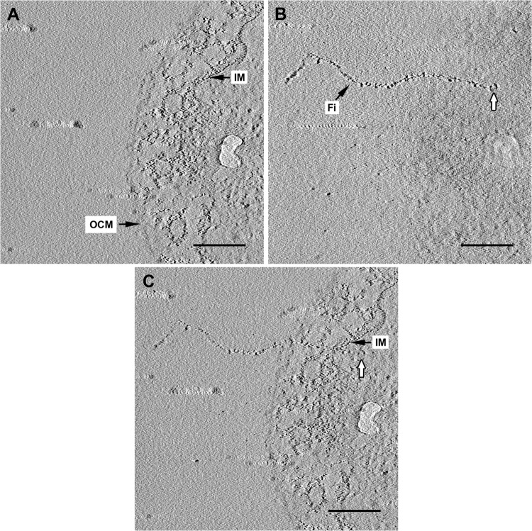
FIG 4.
Electron tomography of an ultrathin section of I. hospitalis. (A, B) Selected slices of the final tomogram after reconstruction of a tilt series of an I. hospitalis cell displaying the membranes (A) and a single fiber, with a knob-like structure at its end inside the cytoplasm (white arrows in panels B and C). (C) After merging both images, the localization of the fiber is more obvious, passing both membranes and being anchored within the cytoplasm. IM, inner membrane; OCM, outer cellular membrane; Fi, fiber. Scale bars, 200 nm.

