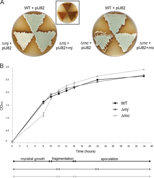
FIG 2.
Phenotypic comparison of wild-type S. venezuelae and the Δrnc and Δrnj mutants. (A) Colony morphologies of wild-type (WT) S. venezuelae and RNase mutants (carrying the integrating plasmid vector pIJ82) and the corresponding complemented strains grown on MYM agar medium for 4 days. The inset image shows the underside of the plate to the left, bearing the rnj mutant and wild-type-complemented mutant strains in the same order and orientation as that for the larger rnj panel (mutant, bottom left; wild type, top; complemented, bottom right), showing relative levels of the brown melanin pigment. (B) Comparison of growth rates and transitions between life cycle stages of wild-type, Δrnj, and Δrnc strains. Cultures were inoculated to an OD600 of 0.05 and incubated with shaking at 30°C for 34 h. The optical density (OD600) was measured at the indicated times, and life cycle stages (indicated below the graph) were assessed using light microscopy. Each OD600 value represents an average from three to four replicates; the standard error for growth density was calculated at each time point.

