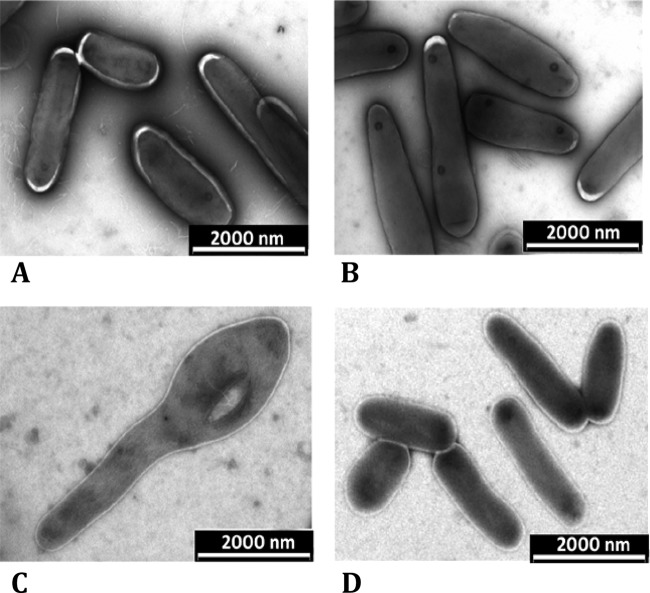
FIG 5.
Electron microscopy of representative stationary-phase wild-type (WT) and mutant (KK104-10) M. smegmatis cells at the end of the growth curve in low-salt LB broth. Shown are the WT without d-glutamate (A) and with 60 mM d-glutamate supplementation (B) and KK104-10 mutants without d-glutamate (C) and with 60 mM d-glutamate supplementation (D).

