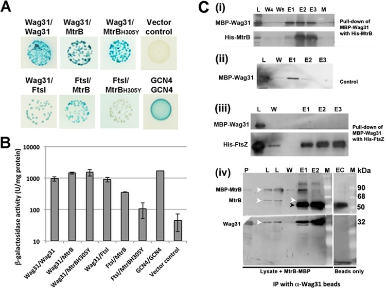
FIG 1.
MtrB interactions with FtsI and Wag31. (A) BACTH analysis of MtrB interactions with FtsI and Wag31. E. coli BTH101 cotransformants with plasmids expressing Wag31/Wag31, Wag31/MtrB, Wag31/MtrBH305Y, MtrB/FtsI, and MtrBH305Y/FtsI were spotted on indicator agar plates as previously described (18), wherein blue and white spots indicate positive and negative interactions, respectively. Cotransformants with GCN4/GCN4 and empty vector/MtrB, representing positive and negative controls, respectively, were also spotted. (B) Recombinant colonies expressing the indicated combinations of proteins were propagated in LB broth, and β-galactosidase activity was measured as described in the text. Data shown are means ± standard deviations from three independent experiments. (C) Pulldown assays. Wag31 interacts with MtrB and not with FtsZ. E. coli lysates with MBP-Wag31 were incubated with His10-MtrBsol (i) or His10-FtsZ (iii) bound to Ni-NTA resin with rocking at 4°C. The Ni-NTA resin was washed five times, and the bound proteins were eluted with buffer containing imidazole (see below and Materials and Methods) (17). MBP-Wag31 was incubated with Ni-NTA (ii) and processed as described for panels i and iii. Various fractions were separated in SDS-polyacrylamide gels and transferred to PVDF membranes, and the proteins were visualized by immunoblotting using anti-MtrB, anti-Wag31, anti-FtsZ, or anti-MBP antibody (see Fig. S2 in the supplemental material). L, load; W, wash; E, elution fractions; W4 and W5, washes 4 and 5, respectively; E1, E2, and E3 are elutions with buffers containing 100 mM, 300 mM, and 1 M imidazole, respectively. For co-IP assays (iv) 1 ml of M. smegmatis lysate mixed with 60 μg of purified MBP-MtrB was incubated overnight with anti-Wag31 antibodies coupled to magnetic beads (BioMag Plus amine particles). The beads were washed five times with IP wash buffer, and the MtrB immunoprecipitates were eluted with 100 mM citrate (pH 3.1). The eluted proteins were neutralized with 1 N NaOH, separated by SDS-PAGE, and visualized following immunoblotting with anti-Wag31 or anti-MtrB antibody. P, pure protein markers for MBP-MtrB and His-Wag31; L, load; W, wash 5; E1 and E2, elutions 1 and 2; EC, elution from buffer control where beads were incubated with buffer instead of lysate. White arrowhead, MBP-MtrB, MtrB, or Wag31; black arrowhead, IgG band. M, molecular mass marker. α, anti.

