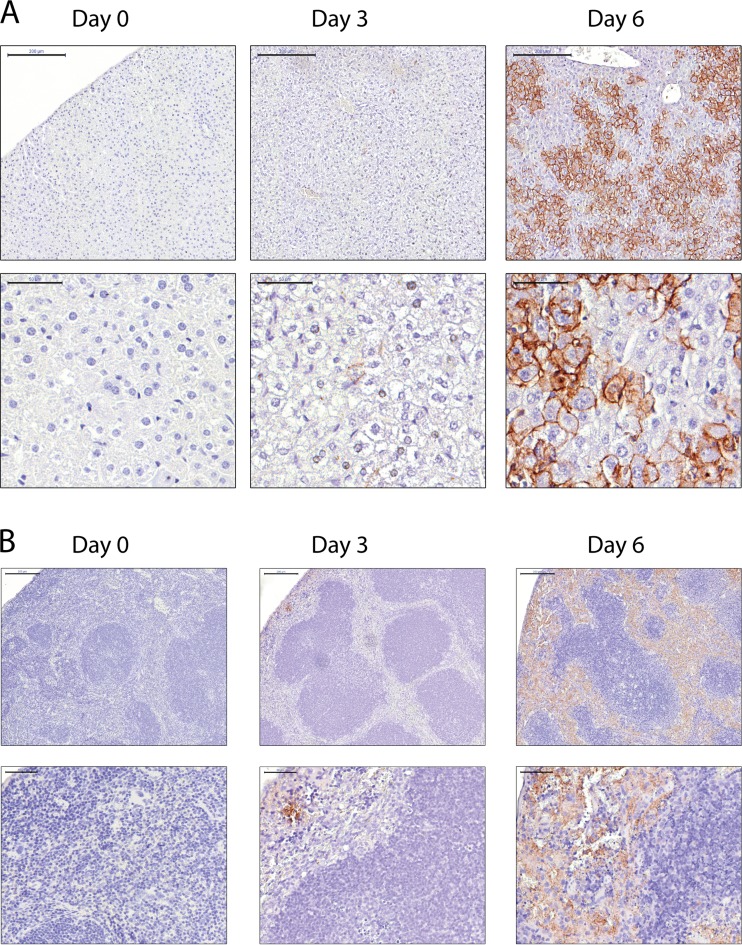
FIG 8.
Immunohistochemistry findings in the livers and spleens of BALB/c mice infected with MARV/Ang-MA. BALB/c mice (n = 4) were infected i.p. with 2,000× LD50 of MARV/Ang-MA. Representative picture of livers (A) and spleens (B), which were collected at the indicated time points and with immunohistochemical staining performed using a monoclonal antibody against the MARV glycoprotein (GP), are shown. Top panels are of lower magnification, and bottom panels are of higher magnification (scale bars, 200 and 50 μm, respectively).

