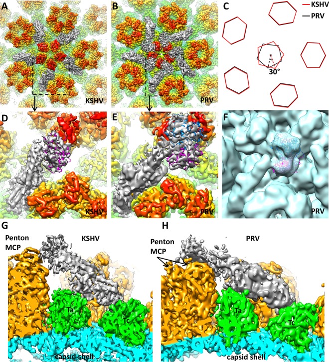
FIG 2.
Comparison of capsid-associated tegument densities in KSHV and pseudorabies virus (PRV). (A and B) Densities around the capsid vertex in the 6-Å KSHV virion reconstruction (A) and 9-Å PRV C-capsid reconstruction (B) (rendered from EMDB-5650; published by Homa et al. [17]). In both structures, capsid proteins are rainbow colored radially and tegument densities are gray, and they are displayed at a lower contour level than that of the capsid because they are weaker than capsid densities. (C) Schematic representation of pentons and periphery hexons in KSHV and PRV. The PRV penton has a 30° clockwise rotation when its periphery hexons are aligned to those of KSHV. (D and E) Zoomed-in views of a single capsid-associated tegument density in KSHV and PRV. Their positions are denoted with dashed squares in panels A and B. Note the slightly different orientations of the tegument density relative to the penton MCP. In KSHV (D), a single copy of the HSV-1 UL25 atomic model (magenta ribbon; PDB entry 2F5U) (39) can be fitted into the tegument density, while in PRV (E) two copies (magenta and blue ribbons) can be accommodated. (F) Two copies of the HSV-1 UL25 atomic model (magenta and blue ribbons) fitted in the tegument density of the PRV virion reconstruction (light blue) (EMDB-5655; Homa et al. [17]). Note the good match between the overall shape of the density map and that of the model. (G and H) Side views of tegument density interacting with penton and triplexes Ta and Tc in KSHV (G) and PRV (H), respectively.

