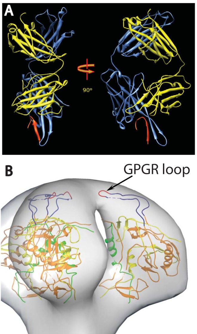
FIG 1.

(A) Ribbon representation of the crystal structure of Fab 447D MAb from PDB 1Q1J (58). The heavy chain is colored cornflower blue and the light chain is colored yellow, with the V3 MN peptide in red. The two views are approximately perpendicular to the pseudo 2-fold axis. (B) View of a plausible spike atomic model showing the rough location of the V3 loop. The 3-fold axis of the spike is approximately vertical in the picture. The coloring scheme is as follows: gp120 residues 90 to 124, green; residues 198 to 396, orange; and residues 410 to 492, yellow. The intact V3 loop, residues 298 to 327, is navy blue, and the GPGR loop at the tip of V3 is red.
