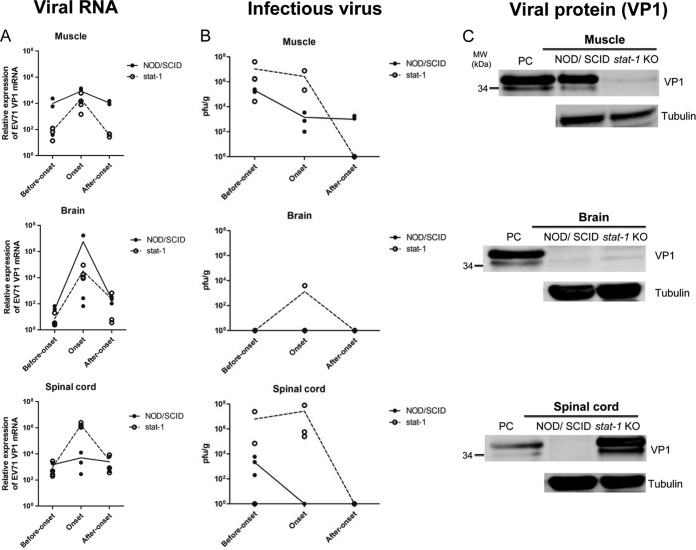
FIG 6.
Side-by-side kinetic comparisons of viral RNA copy numbers, infectivity, and VP1 protein expression in muscle, brain, and spinal cord tissues between NOD/SCID and stat-1 KO mouse models of experimental EV71 infection. One-week-old NOD/SCID and stat-1 KO mice were infected i.p. with EV71 (108 PFU/mouse). Various tissues (muscle, brain, and spinal cord) were collected for analyses of total EV71 VP1 RNA expression by RT-qPCR (A), extracellular infectious virus titers by plaque-forming activity assays (see Materials and Methods) (B), and total VP1 protein expression by Western blotting (C) at three different time points (before disease onset, at onset, and after onset). Lines in the graphs connect the average data points at the different time points. (A) The general trends of the viral RNA profiles are similar for these two mouse models. Viral RNA copy numbers peaked at disease onset in both models. (B) Unlike the viral RNA expression profiles shown in panel A, the infectious titers of EV71 were different in these two models. Infectious EV71 was not detectable by the PFU assay at or after disease onset in the brains or spinal cords of NOD/SCID mice. In contrast, after onset, infectious virus could be easily detected in the muscles of NOD/SCID mice but not in those of stat-1 KO mice. (C) At disease onset, while VP1 protein was detected in the muscles of NOD/SCID mice, it was not detectable in those of stat-1 KO mice. In contrast, while VP1 protein was detected in the spinal cords of stat-1 KO mice, it was not detectable in those of NOD/SCID mice. No VP1 protein was detected in the brain tissue in either model. The lower band of the VP1 doublet could represent a degradation product of the full-length protein. PC, positive control, prepared from the culture medium of EV71-infected RD cells.

