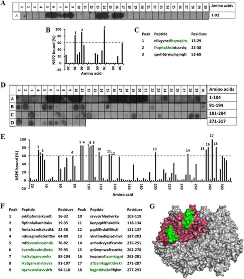
FIG 9.
Mapping the NSP2-NSP6 interaction sites. (A) Autoradiograph of the NSP6 peptide array probed by full-length NSP2. The peptide array consists of spots of 17-residue peptides in the protein sequence, starting from the N terminus (spot A1) and ending with the C-terminal peptide (A30), with the N-terminal residue of the peptide in each spot shifted by 3 residues from the previous spot along the protein sequence. (B) Graph showing the relative intensity (y axis) of each spot (black bars) in the array with its position relative to the protein sequence (x axis). (C) The peptide sequences corresponding to the spots denoted 1 to 3 in panel B, which showed intensities higher than the 60% cutoff. (D) Autoradiograph of the NSP2 peptide array probed by full-length NSP6. The peptide array consists of spots of 17-residue peptides in the protein sequence starting from the N terminus (spot A1) and ending with the C-terminal peptide (D11), with the N-terminal residue of the peptide in each spot shifted by 3 residues from the previous spot along the protein sequence. (E) Graph showing relative intensity (y axis) of each spot (black bars) in the array with its position relative to the protein sequence (x axis). (F) The peptide sequences corresponding to the spots denoted 1 to 18 in panel B, which showed intensities higher than the 60% cutoff. NSP2 residues present in at least two peptides that showed ≥80% binding intensity (green) were mapped onto the known structure of NSP2 (pdb 1L9V). (G) Surface model of the NSP2 octamer (gray) with NSP6-binding residues shown in green on monomer A (pink).

