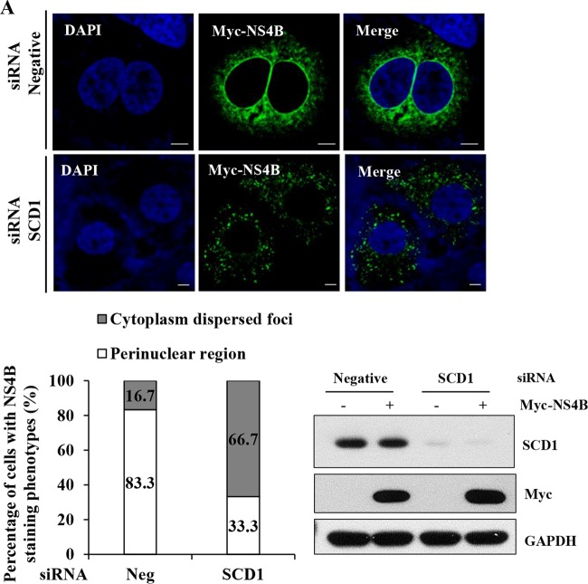
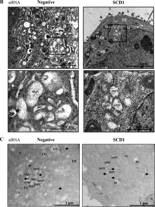
FIG 7.
Knockdown of SCD1 impairs NS4B-induced membrane rearrangement. (A) Huh7.5 cells were transfected with the indicated siRNAs. At 24 h after transfection, cells were further transfected with Myc-tagged NS4B for 48 h. Cells were then fixed and processed for immunofluorescence assay with Myc-tagged NS4B antibody (green). Cells were counterstained with DAPI to label nuclei (blue) (upper panel). Bars, 5 μm. (Lower left panel) NS4B staining pattern of either cytoplasmic dispersed foci or perinuclear large foci was quantitated in 30 randomly selected NS4B-positive cells and presented as percentage. (Lower right panel) Protein expressions were confirmed by immunoblot analysis with the indicated antibodies. (B) Huh7.5 cells were transfected with either negative siRNAs or SCD1 siRNAs. At 48 h after transfection, cells were further transfected with Myc-tagged NS4B expression plasmid for 36 h. Cells were then fixed and processed for electron microscopy analysis. The dark square boxes from upper panels are enlarged with higher magnification and presented in lower panels. N, nucleus; M, mitochondria; ER, endoplasmic reticulum; SMV, single membrane vesicle. Bars, 2 μm (upper) and 500 nm (lower), respectively. (C) Immunogold electron microscopy was performed to verify the presence of NS4B in the membranous web structures as described in Materials and Methods. Arrows indicate immunogold particles of NS4B. N, nucleus; M, mitochondria; ER, endoplasmic reticulum; SMV, single membrane vesicle; LD, lipid droplet. Bars, 1 μm.


