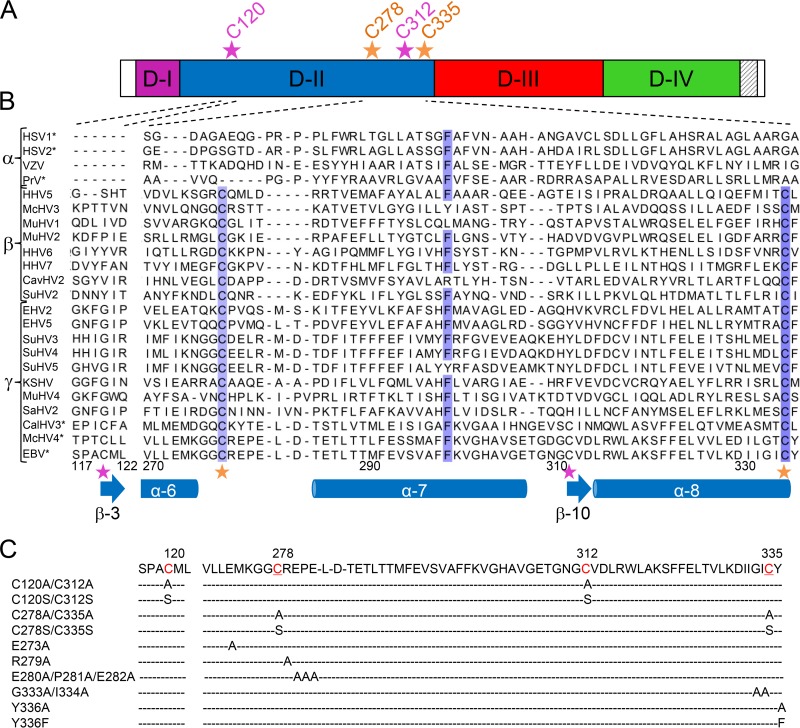
FIG 2.
Site-directed mutagenesis of the DB C120/C312 and C278/C335 of D-II of EBV gH. (A) Schematic overview of EBV gH. The four domains of gH are colored according to the scheme of the ribbon diagram in Fig. 1A, and the transmembrane domain is indicated by gray stripes on a white background. The locations of the DB C120/C312 (light magenta) and C278/C335 (orange) of D-II are indicated by an asterisk and the position of the amino acid. (B) Amino acid alignment of the DBs of D-II of EBV gH is shown with the correlated secondary structures, such as α-helices and β-sheets. Amino acid sequences for several gH homologs of the three subfamilies of the Herpesviridae are shown for a partial D-II, proceeding from amino acid 117 to 122 and 270 to 336 of EBV gH. The locations of the cysteines of the DB C120/C312 (light magenta) and C278/C335 (orange) of D-II are indicated by asterisks. HSV-1 and HSV-2, herpes simplex virus 1 (GenBank accession no. AAG17895.1) and 2 (CAB06746.1), respectively; VZV, varicella-zoster virus (ABF22268.1); PrV, pseudorabies virus (CAA41678.1); HHV5, HHV6, and HHV7, human herpesvirus 5 (CAA00301.1), 6 (CAA58382.1), and 7 (AAB64293.1), respectively; McHV3 and McHV4, macacine herpesvirus 3 (AAP50630.1) and 4 (AAK95464.1), respecitvely; MuHV1, MuHV2, and MuHV4, murid herpesvirus 1 (AAA20190.1), 2 (NP_064175.1), and 4 (NP_044860.1), respectively; CavHV2, caviid herpesvirus 2 (P87730.2); SuHV2, SuHV3, SuHV4, and SuHV5, suid herpesvirus 2 (YP_008492985.1), 3 (AAM22122.1), 4 (AAO12364.1), and 5 (AAO12326.1), respectively; EHV2 and EHV5, equid herpesvirus 2 (NP_042618.1) and 5 (ACY71880.1), respectively; KSHV, Kaposi's sarcoma-associated herpesvirus (ADB08188.1); SaHV2, saimiriine herpesvirus 2 (P16492.1); CalHV3, Callitrichine herpesvirus 3 (AAK38222.1); and EBV, Epstein-Barr virus (P03231.1). The grade of conservation is indicated in blue for the best-conserved amino acids. Analysis was performed with T-COFFEE Expresso (http://tcoffee.crg.cat/apps/tcoffee/do:expresso), which aligns protein sequences using structural information, such as the PDB files (45). The PDB files (PDB entries 3M1C, 2XQY, and 3PHF) are assigned, based on the amino acid sequence, to the corresponding herpesviruses (HSV-1, HSV-2, PrV, CalHV3, McHV4, and EBV), which are labeled with asterisks. The alignment was modified using Jalview (46). (C) The DBs and adjacent amino acids of the conserved DB of D-II of EBV gH were mutated by site-directed mutagenesis. The cysteines of the DBs are indicated in red and by position; in addition, the conserved motif is underlined.

