
FIG 2.
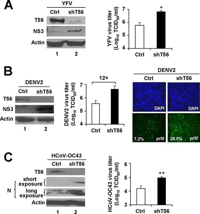
The endogenous TRIM56 protein restricts propagation of YFV, DENV2, and HCoV-OC43. (A) (Left side) Immunoblot analysis of the expression of TRIM56 (using anti-TRIM56 antibody) and YFV NS3 (using anti-YFV NS3 antibody) in HeLa cells stably transduced with either a nontargeting, scrambled control shRNA (Ctrl) or TRIM56 shRNA-094 (shT56) at 72 h after infection with YFV-17D (MOI = 0.5). Actin served as a loading control to demonstrate equal sample loading. (Right side) Culture supernatants of virus-infected cells were harvested at 36 hpi and subjected to TCID50 assay for determination of progeny virus production. (B) (Left side) Immunoblot analysis of TRIM56 and DENV2 NS3 (using anti-YFV NS3) expression in HeLa-Ctrl and HeLa-shT56-094 cells at 60 h after infection with DENV2-16681 (MOI = 1.5). (Middle) Progeny virus titers in culture supernatants of the DENV2-infected cells (MOI = 0.5, 60 hpi). Note that HeLa-Ctrl cells produced 12-fold-fewer progeny viruses than HeLa-shT56-094 cells. (Right side) Immunostaining of DENV2 prM expression (using DENV 2H2 hybridoma supernatant) in HeLa-Ctrl and HeLa-shT56-094 cells at 60 h after infection with DENV2-16681 (MOI = 0.5). The mean percentage of cells with discernible prM expression was quantified from three independent images and is shown at the lower left corner of each prM panel. (C) (Left side) Immunoblot analysis of TRIM56 and N protein (N) (using anti-HcoV OC43 N) expression in HeLa-Ctrl and HeLa-shT56-094 cells at 48 h after infection with HCoV-OC43 (MOI = 0.01). (Right side) Progeny virus titers in culture supernatants of virus-infected cells. Single and double asterisks indicate that statistical differences exist between Ctrl and shT56 cells with P values of <0.05 and <0.01, respectively.