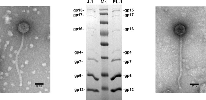
FIG 3.
Analysis and functional assignment of J-1 and PL-1 structural proteins. Illustrated are electron micrographs of J-1 (left) and PL-1 (right) phage particles and SDS gel electrophoresis of virion proteins showing the predicted gene products. Molecular mass markers (Mk) are, from top to bottom, 170, 130, 100, 70, 55, 40, 35, and 25 kDa.

